Aboudoulatif Diallo1 ![]() ,
Paolo Darre2,
Kossi Metowogo3,
Povi Lawson-evi3,
Divakar Selva4,
Yao Potchoo1,
Edmond Creppy5,
Muthiah Ramanathan4
,
Paolo Darre2,
Kossi Metowogo3,
Povi Lawson-evi3,
Divakar Selva4,
Yao Potchoo1,
Edmond Creppy5,
Muthiah Ramanathan4
For correspondence:- Aboudoulatif Diallo Email: aboudoulatif@gmail.com Tel:+22890113723
Received: 15 October 2015 Accepted: 12 January 2016 Published: 28 February 2016
Citation: Diallo A, Darre P, Metowogo K, Lawson-evi P, Selva D, Potchoo Y, et al. Fetal toxicity and cytotoxicity of Lannea kerstingii Engl and Krause stem bark (Anacardiaceae). Trop J Pharm Res 2016; 15(2):253-258 doi: 10.4314/tjpr.v15i2.5
© 2016 The authors.
This is an Open Access article that uses a funding model which does not charge readers or their institutions for access and distributed under the terms of the Creative Commons Attribution License (http://creativecommons.org/licenses/by/4.0) and the Budapest Open Access Initiative (http://www.budapestopenaccessinitiative.org/read), which permit unrestricted use, distribution, and reproduction in any medium, provided the original work is properly credited..
Purpose: To evaluate the fetal toxicity and cytotoxicity of L. kerstingii in pregnant rats exposed in the organogenic period.
Methods: Mated female rats were randomly assigned to three experimental groups of 8 animals each. Pregnant rats received orally 500 or 1000 mg/kg of 50 % hydroalcohol extract of L. kerstingii, daily from the 17th to the 20th day of gestation. On day 21 of pregnancy, the females were sacrificed. Laparotomy was performed and uterine horns were removed. The number of implants, resorptions, dead and live fetuses were then recorded. The ovaries were also observed and corpora lutea were counted. The cytotoxic effect of L. kerstingii hydroalcohol extract was evaluated on Caco-2 cell lines using MTT (3-(4, 5-dimetylthiazol-2-yl)-2, 5- diphenyltetrazolium bromide) and neutral red uptake assay.
Results: No visible signs of toxicity were observed in the female rats and their pups through-out the study period. However, L. kerstingii (500 and 1000 mg/kg) caused a significant de-crease (p < 0.01) in fetal weight compared with control. With regard to implantation, resorption and mortality, there was no significant difference between groups. L. kerstingii hydroal-cohol extract (IC50, 29 µg/mL) was more cytotoxic than the aqueous extract (IC50, 141 µg/mL).
Conclusion: The administration of hydroalcohol extract of L. kerstingii to female rats in late pregnancy is toxic to the fetus.
Introduction
The use of herbal plants as natural remedies, functional foods, and dietary supplements for health care has been increasing in the world. Market estimates suggest that the rate of growth in sales of traditional medicinal products in recent years is between 5 and 18 % per annum [1].
One of these traditional medicines is Lannea kerstingii Engl. and K. Krause (Anacardiaceae). L. kerstingii stem bark decoction gives a red color. Many women use it during pregnancy or during lactation in Togo to treat anaemia and malaria [2]. In West African countries such as Ivory coast, L. kerstingii stem bark and root are consumed as traditional remedies for the treatment of diarrhoea, gastritis, rheumatic, sterility, scorbut, scurvy, epilepsy and intestinal helminthiasis [3,4]. The fruit of L. kerstingii is eaten raw in Guinean pre-forest savannas of Ivory Coast [5]. In Benin, L. kerstingii leaves are used in the treatment of Buruli ulcer [6].
Pharmacological studies of L. kerstingii extracts have revealed several properties such as antihelmintic, antimicrobial, trypanocidal and acetylcholinesterase inhibitory properties [7,8]. Our previous study has shown the antioxidant effect of L. kerstingii stem bark hydroalcohol extract using appropriate in vivo and in vitro [3]. During a 28 and 60 days subchronic test, L. kerstingii at 1000 mg/kg increased significantly the relative weight of the spleen, decreased significantly (the increment of body weight and induced 50 % of rats’ death with an accumulation of gas in the digestive tract [9,10]. Body weight changes are an indicator of adverse side effects, as the animals that survive cannot lose more than 10 % of the initial body weight [11]. Apart from the presence of gas in the gastrointestinal tract, L. kerstingii decreased significantly the daily food intake and rats’ intestinal motility.
Based on the knowledge that toxicity often occurs during pregnancy and may have different effects on embryo development depending on the conceptus phase and the maternal conditions [12] and that L. kerstingii is widely used in folk medicine [2-8], the aim of the present investigation is to evaluate the effect of L. kerstingii on pregnant rats exposed to this plant in the organogenic period.
Methods
The study was conducted following an approved animal use protocol from the Institutional Ethical Committee for Teaching and Research (ref no. CNCB- CEER 2801/2010). Animal care and handling conducted conformed to accepted guidelines [13].
Plant materials
Lannea kerstingii stem bark was collected at Bagbe (Togo) in July 2012. It was identified by Prof Kouami Kokou from the Botany Department of University of Lome (Togo) and a voucher specimen (N0 10553 Akpagana) was kept in the herbarium of the Laboratory of Botany and Plant Ecology (Faculty of Science/University of Lome.
Preparation of hydroalcohol extract
The stem bark was washed in running water, then dried and ground to powder. The powder was soaked in ethanol-water (50-50: v/v) for 72 h with manual discontinue agitation. The solution was filtered and evaporated using a rotary evaporator (yield: 12.34 %). The study was conducted in Animal Physiology Department, Faculty of Sciences, University of Lome, Togo.
Animals
Female Wistar rat (150 - 200 g), provided by the Department of Animal Physiology of University of Lome (Togo) were used. They were housed in a standard environmental condition and fed with rodent standard diets and water ad libitum.
Fetal toxicity test
Female rats were mated with fertile males and the presence of spermatozoa in the vagina or seminal plug was considered as day 1 of pregnancy. The mated females were randomly as-signed to three experimental groups of 8 animals each Group 1 received 10 ml/kg of distilled water and served as control. Group 2 and 3 received L. kerstingii hydroalcohol extract at 500 mg/kg body wt. and 1000 mg/kg body wt. respectively from the 17th to the 20th day of gestation. Doses of 500 and 1000 mg/kg are therapeutic doses used in our previous study [3]. Animals were observed at least twice daily for morbidity and mortality. Body weight of animals was evaluated daily. On day 21 of pregnancy, the females were sacrificed. Laparotomy was performed and uterine horns were removed. The number of implants, resorptions, and dead and live fetuses were then recorded. The ovaries were also observed and the corpora lutea were counted.
To study the reproductive capacity of the female rats, the implantation (number of implantations/number of corpora lutea x 100), resorption (number of resorptions/number of implantations x 100) and mortality indices (number of dead fetuses/number of fetuses x 100) were calculated. The fetuses were weighted and examined for external macroscopic malformation
In vitro toxicity
Caco-2 cells (human colon cancer line) were obtained from Dr. Jing Yu, (Tuffs School of Medicine, Medford, MA, USA). The cells were routinely cultured in a humidified 5 % CO2 – 95 % air mixture at 37 °C and were grown in DMEM medium (Sigma, France), supplemented with 10 % foetal bovine serum, 8 mM L-glutamine, penicillin (100 UI/mL) and streptomycin (100 µg/mL).
Neutral red uptake assay
Caco-2 cells were seeded in 96-wells microplates (10000 cells/200 µL/well) and routinely cultured in a humidified incubator for 24 h. Cells were maintained in culture and exposed to plant extracts over a range of concentrations (10-500 µg/mL). After 72 h exposure to extracts, neutral red uptake test (NR) was performed according to the procedure described by Yusup et al [14]. At the end of the treatment (72 h), the medium with or without extracts was discarded, and 200 µL of freshly prepared neutral red solution (50 µg/mL) was added to each well. Cells were then re-incubated for an additional 4 h at 37 °C. Thereafter, the cells were carefully washed twice with 200 µL of PBS to eliminate extracellular neutral red. The incorporated dye was eluted from the cells by adding 200 µL elution medium (50 % ethanol supplemented with 1 % acetic acid, v/v) to each well followed by gentle shaking of the microplate for 15 min. The plates were then read at 540 nm using a microplate reader (Dynatech MR 4000, Dynatech, Boston, MA, USA). Surviving cells in treated wells were expressed as percentage of control wells. The IC50 (50 % viability inhibitory effect) was determined graphically and expressed in µg/mL.
MTT assay
This test was carried out according to the method described by Kouadio et al [15]. Caco-2 cells were seeded in 96-well microplates (10000 cells/µL/well) and routinely cultured in a humidified incubator for 24 h. Cell culture media were removed and extracts were added in concentration ranging from 10 to 500 µg/mL. Cells were then incubated for 72 h. In this test, a control group (DMEM without extract) and a blank group (without cells or medium) were also included. The medium with or without extract was then discarded, and 100 µL of tetrazolium salt MTT (3-[4, 5-dimethylthiazol-2-yl]-2,5-diphenyl tetrazolium bromide) solution (0.5 mg/mL in DMEM or RPMI) were added to each well. Cells were re-incubated for an additional 2 h; 100 µL of 10 % SDS in 0.01 M HCl were added to each well to dissolve the formazan crystals. The plates were then read on a microplate reader (DYNATECH MR 4000, Dynatech, Boston, MA, USA) at 560 nm. Four wells were used for each concentration. Surviving cells in treated wells were expressed as percentage of control wells. The IC50 (50 % viability inhibitory effect) was determined graphically and expressed in µg/mL.
Induction of lipid peroxidation in Caco-2 cells
Caco-2 cells were cultured (1 x 10⁵ cell/mL) in 24-well non-coated microplates, for 19 h at 37 °C. L. kerstingii hydroalcohol (50 %) and aqueous extracts (500 μg/mL) and H2O2 (10 mM) prepared in cells culture medium were added. After 24 and 48 h of contact, the supernatant was removed. Then cells were trypsinised, centrifuged and resuspended in SET buffer (0.1 M NaCl, 20 mM EDTA, 50 mM Tris-HCl, pH 8.0) for the determination of MDA level.
Determination of MDA-TBA adduct
Lipid peroxidation was measured by quantification of MDA-TBA adducts formed during incubation, as previously described by Yusup et al [14] and related to the protein content of Caco-2 cells homogenates. The protein content was determined through the use of Bradford method [16].
Statistical analysis
The results are expressed as mean ± standard error of the mean (SEM). Statistical analysis was performed by one-way analysis of variance (ANOVA) with Tukey test to evaluate significant differences between groups. P < 0.05 was considered statistically significant. Statistical analysis was carried out using Instat Statistical package (Graph Pad software, Inc, USA).
Results
Fetal toxicity
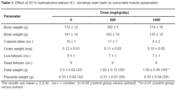
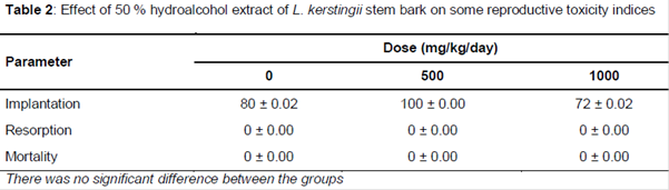
All rats from control and treated group survived throughout the study period. No visible signs of treatment such as changes in respiratory, circulatory, autonomic and central nervous system, behavioral pattern were observed in females and their pups throughout the study period. However, L. kerstingii (500 and 1000 mg/kg) caused a significant decrease (p < 0.01) of fetal weight compared with the control (). For the implantation, resorption and mortality there was no significant difference between groups ().
Cytotoxicity
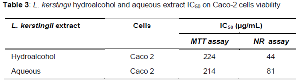
The IC50 obtained with the neutral red and MTT assay are presented in . On Caco-2 cells, L. kerstingii hydroalcohol extract and its aqueous extract showed a similar toxicity ex-hibiting a close IC50 (102 and 104 µg/mL respectively) but with the neutral red assay, L. kerstingii hydroalcohol extract (IC50, 29 µg/mL was more toxic () than its aqueous ex-tract (IC50: 141 µg/mL).
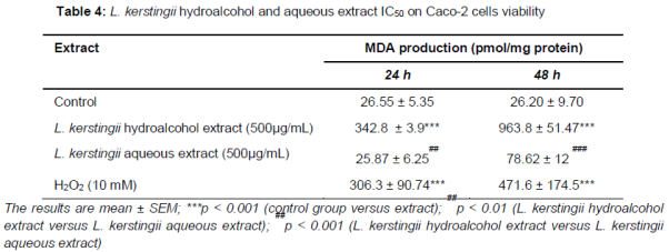
The hydroalcohol extract of L. kerstingii (500 µg/mL) and its aqueous extract increased significantly MDA levels. As for the cytotoxicity test, the hydroalcohol extract caused a larger increase of MDA levels than the aqueous extract and the positive control ().
Discussion
When testing possible fetal toxic effects of a specific substance, it is necessary to establish if these effects are due to direct action on the fetus or an indirect action through the maternal organism that could secondarily interfere with the fetus. There are many ways of evaluating maternal toxicity. The clinical criteria suggested by Khera [17] and by Mason and Kang [11] for the evaluation of maternal toxicity are food intake, body weight, piloerection, locomotor activity, diarrhoea and vaginal bleeding.
L. kerstingii has induced fetotoxicity at 1000 mg/kg. Fetal toxicity may be due to direct or indirect action. For example, a decrease in blood sugar or anorexia can decrease fetal weight [18]. Indeed, the fetus requires energy and nutrients for development. A decrease in blood sugar or in energy can logically lead to reduced fetal weight. In this study we have evaluated the hypoglycemic effect of L. kerstingii at 500 and 1000 mg/kg. The results show that L. kerstingii does not decrease the blood glucose level (data not shown). In our preliminary studies, L. kerstingii administered for 28 and 60 days, caused a significant decrease in the weight of the Wistar rats and decreased the quantity of food consumed [9,10]. This toxicity of L. kerstingii characterized by anorexia and a decrease of rat’s weights can explain the decreased of the fetus weight [9,10]. The lipid peroxidation test has shown that the hydroalcohol extract of L. kerstingii was responsible of a strong oxidative stress. Several authors believe that oxidative stress may cause fetal toxicity [11-17].
Oxygen radicals or reactive oxygen species (ROS) act as primary or secondary messengers to promote cell growth or death. Many studies demonstrate an important direct role of ROS in development because redox status regulates key transcription factors that influence cell signaling pathways involved in proliferation, differentiation, and apoptosis [11,18]. Therefore, oxidative stress can alter many important reactions that affect embryonic or fetus development both positively and negatively [17,18]. It is known that the implantation index, that correlates the corpora lutea with the number of implantations in the uterine horn, is an indicator of the reproductive capacity success [19]. The data obtained in this study indicate that the number of blastocysts implanted was similar between control and treated groups. The resorption index indicates the failure in the progress of the embryo development [19]. As the resorption index was similar in all experimental groups, it may be assumed that experimental group failures in the progress of embryo development are similar.
Conclusion
Administration of hydroalcohol extract of L. kerstingii to female rats in late pregnancy is toxic to the fetus as it causes weight reduction in fetal weight. The fetal toxicity may be due to the oxidative stress induced by the components of the plant extract.
References
Archives


News Updates